Eye Anatomy
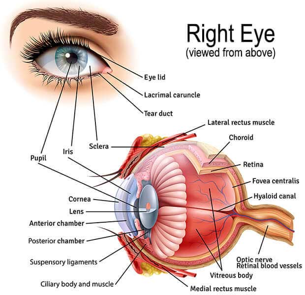
Anterior Chamber
Fluid-filled space inside the eye between the iris and the innermost corneal surface.
Anterior Chamber Angle
Junction of the front surface of the iris and the back surface of the cornea, where aqueous fluid filters out of the eye.
Choroid
The vascular layer of the eye lying between the retina and sclera. This layer furnishes nourishment to outer layers of the retina.
Ciliary Body
A ring of tissue inside the eye composed of ciliary muscle, which is involved in lens focusing and control of the intraocular pressure. There are also ciliary processes that produce aqueous.
Cornea
The transparent front segment of the eye that covers the iris, pupil and anterior chamber, and provides most of the eye’s optical power.
Iris
Colored tissue lying behind the cornea that gives color to the eye (e.g., blue eyes, brown eyes) and controls the amount of light entering the eye by varying size of the black pupillary opening.
Lens
Transparent, intraocular tissue that helps bring rays of light to focus on the retina. A cataract is a lens that has become cloudy or opaque.
Macula
The centralized area of the retina responsible for acute central vision necessary for reading and detail work.
Optic Nerve
The primary sensory nerve of the eye. It carries impulses for sight from the retina to the brain.
Pupil
A black circular opening in the center of the iris that regulates the amount of light that enters the eye.
Retina
The part of the eye that converts images from the eye’s optical system into electrical impulses sent along the optic nerve for transmission to the brain. The retina lines the rear two-thirds of the eye and consists of layers that include rods and cones. This part of the eye can be compared to film in a camera. This part of the eye is directly affected when Macular Degeneration, Diabetic Retinopathy, Retinal Tears & Detachments, and Retinal Vascular Disease occur.
Sclera
The white protective outer layer of the eye.
Vitreous
Transparent, colorless, gelatinous material that fills the rear two-thirds of the interior of the eyeball, between the lens and the retina.
Zonules
Fibers that suspend the lens from the ciliary body and hold it in position.