Posterior Vitreous Detachment
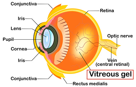
The main cavity of the eye is filled with a gel-like clear substance called the vitreous gel or vitreous humor. As we age, that gel becomes more liquid, losing its rigidity. At some point, enough rigidity is lost to cause the back layer of the vitreous to separate from the retina.
This process is called a Posterior Vitreous Detachment (PVD) or Posterior Vitreous Separation (PVS). While it sounds dramatic, it is part of the natural aging process. In most patients, this separation happens without adverse consequence. However, in some patients, the separation of the vitreous gel can induce a tear in the light-sensing layer of the inside of the eyeball – the retina. This retinal tear is an emergent condition and can lead to retinal detachment if untreated.
The symptoms of a PVD can include:
- Sudden onset of floaters in one eye: black dots or “spider-webs” that move in the field of vision
- Sudden onset of flashes of light in one eye
- A gray or black cloud obscuring a part of the vision
These can be intermittent, but typically, there is an abrupt change. Unfortunately, without a proper dilated retinal examination, it is not possible to determine if there is a normal age-related PVD alone or if there are underlying retinal tears.
For this reason, if you have sudden onset of the symptoms listed above, contact our offices. Our staff will discuss your symptoms, and if appropriate, will promptly get you scheduled with one of our specialized, experienced physicians. CFR physicians do a detailed dilated examination of both retinas and can immediately treat any retinal tears or retinal detachments.
Untreated or unaddressed, the above symptoms can lead to conditions that can be permanently vision-threatening. Contact our office or your eye care professional immediately if you note these symptoms.