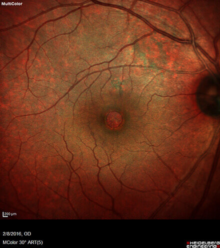
Macular Hole
The macula is the portion of the retina that accounts for central vision. During the separation of the vitreous gel during a posterior vitreous detachment, vitreomacular traction may lead to the formation of a full thickness break (opening) in the macula.
Patients may experience a central loss of vision, scotoma (blind spot), or metamorphopsia (distortion).
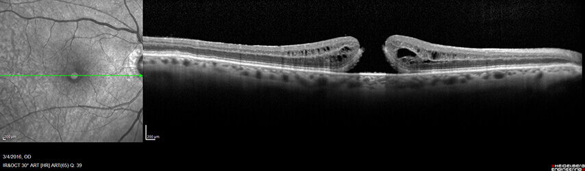
Clinical exam and optical coherence tomography (OCT) are useful for staging and following the response to treatment with macular holes.
Treatment may depend on the specific stage of macular hole and may involve close observation or surgery.
Pars plana vitrectomy is commonly performed to treat macular hole. At the end of surgery, the eye may be filled with a self-dissipating gas which may require special positioning to close the macular hole.
The surgeons at Central Florida Retina are fellowship trained retina specialists practicing the latest techniques for macular hole repair. Should you be experiencing any of these symptoms or would like to request a consultation, please contact (800) 255-7188.